

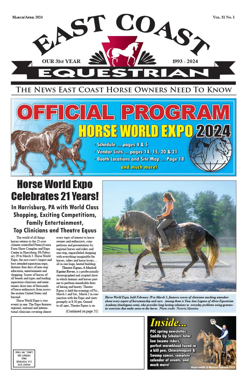
Metabolic diseases in horses was the subject of a presentation at Penn Vet New Bolton Center on January 8 by Olivia Schroeder VMD, DAVBP and a member of the Boucher Field Service at Penn Vets New Bolton Center. The lecture was part of the First Tuesday Lecture series at New Bolton Center, offering the public open lectures on equine topics, at no charge, the first Tuesday of each month.
Metabolic Disease: A Growing Concern
Metabolic diseases in horses, said Dr. Schroeder, are becoming more common. Metabolism has to do with the biochemical processes occurring in every living organism, and is often used to refer specifically to the breakdown of food and its transformation into energy. A recent study from southwestern Virginia investigated the body condition of 300 horses and found that 51 percent of these horses were overweight or obese. While nobody likes to look at an underweight horse, an overweight body condition can be just as dangerous. It puts a strain on every system in the horses body, including the heart, lungs, and especially the musculoskeletal system, worsening clinical signs associated with arthritis and laminitis.
The two main metabolic diseases found in horses are Equine Metabolic Syndrome (EMS) and Pituitary Pars Intermedia Dysfunction (PPID, or Equine Cushings disease). EMS affects primarily younger horses and is characterized by obesity or regional adiposity (fatty deposits), insulin resistance, and laminitis (either clinical or subclinical, that is, having no discernable symptoms). PPID is seen in older horses with classical clinical signs including hirsutism (an excessively thick, long and sometimes curly hair coat that is often slow to shed in the spring), a loss of muscle along the top line, and laminitis.
EMS: How Do You Know?
EMS can be seen in every breed, but ponies, Miniature horses, Morgans, Paso Finos, Arabians, Saddlebreds, and Warmbloods appear to be more susceptible. Most clinical cases are diagnosed between 5 and 15 years of age. There may be a genetic factor that predisposes a horse to developing EMS, but this warrants further investigation. One of the main clinical signs of EMS is regional adiposity, in the area of the nuchal ligament (or the crest of the neck), at the tail head, behind the shoulder, or around the sheath or mammary gland.
So why do our horses develop EMS, when horses in the wild do just fine? While horses under feral conditions will gain weight in the summer when food is plentiful and then lose it in the winter when food is sparse, we are now able to make food plentiful year-round. As a result, horses that would normally lose weight in the winter are able to maintain their body weight through the winter--and then add to it over the summer.
The diagnosis of EMS can be complex and challenging. It should start with a complete history including a thorough examination of the feeding regimen. A physical examination should be performed to look for evidence of regional adiposity and obesity and to examine all other body systems. Blood tests for diagnosis can be divided into resting tests and dynamic tests. Resting tests consist of measuring insulin and glucose concentrations in the blood after a period of fasting. Dynamic testing involves taking a resting blood sample, and then administering either glucose or a combination of glucose and insulin in order to measure the glucose and insulin response.
Diet, Drugs and Exercise
Once a horse is diagnosed with EMS, the primary method of management should be dietary changes. Starches and calories should be reduced to encourage weight loss in obese horses. Ideally, the diet should consist of forage with a vitamin and mineral supplement, rather than pasture grasses. While horses were designed to eat and utilize the energy in pasture grasses, these grasses provide calorie content and starch level that cannot be measured. In horses with confirmed EMS, forage should contain less than 10 percent of dry matter as non-structural carbohydrates. If the carbohydrate content is greater than 10 percent, hay can be soaked in water for 60 minutes to lower the carbohydrate content. Feed should be weighed so amounts being fed are uniform. A digital fish scale is an excellent tool to have around the barn, and will provide a digital read-out of the exact weight of each flake of hay or scoop of feed.
Until insulin sensitivity has improved and the body condition score has decreased, pasture access should be eliminated. Once the horse can be reintroduced to the pasture, grazing should be limited to times when starch content is low, such as early morning. Grazing for fewer than one to two hours a day is recommended, as any more than that can precipitate events leading to laminitis. A grazing muzzle may be worn, or pasture size may be decreased by a fence. If the horse will not wear a muzzle or a small pasture is not available, a dirt paddock may be necessary in order to eliminate any source of starch from the diet.
As is true with humans, it is very difficult for a horse or pony to lose weight without the dreaded combination of diet and exercise. As long as the horse or pony is sound and able to exercise, it is recommended that an exercise regimen be instituted to encourage weight loss. In people, an exercise regimen of 200 minutes per week has been shown to increase insulin sensitivity. There is no definitive recommendation for horses, but in general, two to three sessions per week for 20-30 minutes is a good place to start, with a slow increase in the duration of exercise and the number of sessions per week.
Drugs are the last line in management of horses with EMS. Levothyroxine sodium (Thyro-L) has been shown to increase insulin sensitivity and promote weight loss when given in very high doses for short periods of time. It is a common misconception that this drug is used because horses with EMS also have a problem with their thyroid gland. In reality, the drug is used for its ability to improve insulin sensitivity and promote weight loss. Once the desired weight is achieved, the drug should be given in decreasing levels and then discontinued. It is important that this drug should not be used without also changing the diet, as it can cause horses to become hyperphagic (have an increased appetite) and eat more when on pasture.
Metformin is another drug used in the management of EMS. It improves insulin sensitivity at the level of the individual body tissues and also decreases the production of glucose by the liver. Studies have shown varied responses to metformin, with some studies reporting success and others reporting failure of the drug to reduce insulin concentrations. Other studies have shown questionable absorption of metformin when administered orally, especially with feed, leading to the suspicion that the drug may not achieve high enough levels to become effective. Currently the drug is recommended in some refractory cases of EMS, but further studies are necessary to determine the appropriate dose.
The most important elements of any weight loss program are careful monitoring of progress and long patience. It often takes several months for a horse to lose the desired amount of weight; changes will not be seen immediately. A weight tape should be used to monitor the progress. The same weight tape used every time in the same manner can result in an accurate measurement of how much weight the horse has lost. As a rule of thumb, an average-sized horse must lose approximately 50 pounds in order to achieve a decrease of one body condition score.
Cushings: The What, The Why
Pituitary pars intermedia dysfunction (PPID), also known as Equine Cushings disease, is the other common metabolic disease of horses. A recent UK study based on a resting blood test found that 21 percent of horses 15 years of age or older had PPID. Clinical signs are typically first recognized in horses 18-20 years old, with signs rarely recognized in horses less than 10 years of age. Certain breeds appear to be predisposed to developing PPID, especially ponies and Morgans, but findings have been varied in different studies.
The number one clinical sign of horses with PPID is hirsutism, along with muscle loss along the topline, increased drinking and urination, chronic laminitis, frequent foot abscesses, infections that are difficult to treat, abnormal fat distribution, and (rarely) seizures or blindness.
PPID is caused by a lack of dopamine input to the pars intermedia of the pituitary gland. The pars intermedia is primarily responsible for producing pro-opiomelanocortin (POMC), a compound which is then broken down into many smaller compounds. One of these is ACTH, which is ultimately responsible for stimulating the production of cortisol, a major stress hormone in most mammals. In a normal horse, most of the ACTH that is produced in the pars intermedia is controlled by the production of dopamine. In a horse with PPID, there is a lack of dopamine, so the production of ACTH goes into overdrive.
Diagnosis of PPID can be challenging. Examination of the hair coat is 86 percent accurate and is currently better than any of the blood-based diagnostic tests we have available. As is true with EMS diagnostic tests, PPID blood tests can be divided into resting tests and dynamic tests. Because we are still searching for the perfect test, many options are available. In addition, due to the seasonal variation in the success of blood testing--with fall being the most difficult time to test horses for PPID--many labs are developing new seasonal reference values.
The baseline ACTH test is the easiest test to perform, as it does not require administration of any other substance. In horses with PPID, blood levels of ACTH will be abnormally elevated. This is the blood test that is most susceptible to seasonal changes, because ACTH levels increase in the fall in healthy horses. Recent research has shown that in horses with PPID, this fall elevation of ACTH levels is even greater. This test can still be used, as long as the lab analyzing the blood sample uses appropriate seasonal ranges. There is evidence that stress or pain may increase ACTH levels, complicating their interpretation. Because of this, if there is a high suspicion that a horse has PPID, but the horse is currently suffering from a laminitis flare-up or a foot abscess, treatment may be instituted and testing may be delayed until the horse is more comfortable.
The dexamethasone suppression test (DST) is a dynamic test that involves administration of a dose of dexamethasone, measuring blood cortisol levels before and 19-24 hours after administration. A normal horse will show a decrease in cortisol after administration of dexamethasone. In contrast, in a horse suffering from PPID, the cortisol levels will remain elevated.
The thyrotropin releasing hormone (TRH) stimulation test is another dynamic test that can be used to diagnose PPID. Normal horses will not produce increased levels of ACTH and cortisol in response to an injection of TRH, whereas horses with PPID will have an increase in the level of ACTH (and cortisol). This test is very promising, as it is both safe and quick to perform. As opposed to the DST, only one farm visit is required, and there is little to no risk of laminitis. Additionally, it has the advantage of being able to detect horses in early stages of the disease. However, there is currently no veterinary formulation of TRH, complicating performance of this test.
The domperidone stimulation test can also be used, but further investigation is required to understand how the time of year affects the results, and to validate the test. This test is useful because domperidone is a drug that is already licensed for use in horses and is quite safe.
Tough to ID, Easy to Treat
While diagnosis of PPID may be difficult, treatment is fairly straightforward. The main treatment for PPID is an FDA-approved formulation of pergolide, which is a dopamine agonist. It acts on the nerves in the brain to cause them to release dopamine, which then acts on the pars intermedia to decrease blood levels of ACTH and other hormones.
Pergolide helps treat many of the symptoms of PPID, such as the long haircoat and loss of muscle tone. However, we cannot guarantee that the drug will prevent the development of laminitis. There are few side effects associated with pergolide, the most common of which is a loss of appetite. This can be alleviated by decreasing the dose and then slowly increasing over time until the desired dose is achieved. Treatment with pergolide is lifelong, and over time, an increase in dose may be necessary due to progression of the disease. Other treatments may be used in conjunction with pergolide, but there is no one treatment that is able to replace pergolide completely.
Medication is only one part of the management of PPID. These horses have other management aspects that should be addressed. Because they may take their time shedding out in the spring, clipping a horse with PPID may help prevent overheating and electrolyte imbalances caused by excessive sweating. Because the feet of horses with PPID can slowly deteriorate due to changes in the internal structures, regular hoof trimming and farrier care are essential to maintain the integrity of the hoof wall and sole. In addition, these horses have altered immune systems, and therefore they may have a decreased resistance to parasites. It is important that an annual fecal egg count be performed to determine each individuals worm burden. Feed should be selected carefully to provide adequate calories for maintaining weight without providing excess calories from starches. High-fat feeds are most helpful, so supplementation with corn oil or beet pulp may be necessary. Older horses in general need more frequent dental care, a fact even more true for horses with PPID, as they are more prone to developing sinus and tooth infections. A veterinarian should examine your horses teeth at least once a year to detect any problems early.
Horses with PPID are living longer and better lives, thanks to earlier detection and better treatment options. As a result, we may be able to help manage and prevent some of the devastating complications of the disease.